Loculated Pleural Effusion Radiology Ct - Figure 1 From Factors Influencing Residual Pleural Opacity In Tuberculous Pleural Effusion Semantic Scholar : Pleural effusion, small to moderate 1 of 5 70:. About 75 ml are required to blunt the posterior costophrenic sulcus (seen on the lateral view) and about as the subpulmonic effusion grows in size, it first fills and thus blunts the posterior costophrenic sulcus, visible on the lateral chest. The lungs and the chest cavity both have a lining that consists of pleura, which is a thin membrane. A pleural effusion represents the disruption of the normal mechanisms of formation and drainage of fluid from the pleural space. Obliteration of left costophrenic angle with a wide pleural based dome shaped opacity projecting into the lung noted tracking along the cardiophrenic angle and lateral chest wall suggestive of loculated pleural effusion, however the. Fundamentally a pleural effusion refers to the collection of fluid between the parietal and visceral pleura.
Images of pleural radiology effusion are shown below. Pleural effusion is a condition in which excess fluid builds around the lung. However, patients can also have neutrophilic loculated tpe, although little data are available concerning the incidence and characteristics of this form of tpe. Differentiate from an elevated hemidiaphragm. Large, loculated pleural effusion 2 of 3 68:
Figure 2 From Multiloculated Pleural Effusion Detected By Ultrasound Only In A Critically Ill Patient Semantic Scholar from d3i71xaburhd42.cloudfront.net The lack of specificity is mainly due to the limitations of the imaging modality. Pleural effusion is a condition in which excess fluid builds around the lung. Pleural effusion refers to a buildup of fluid in the space between the lungs and the chest cavity. Learn about pleural effusion including causes of pleural effusion. Pleural effusion, the pathological accumulation of fluid in the pleural space, is very common. Under normal conditions, pleural fluid is secreted by the parietal pleural capillaries at a rate of 0.01 millilitre per kilogram weight per hour. Large, loculated pleural effusion 2 of 3 68: Loculated effusions are collections of fluid trapped by pleural adhesions or within pulmonary fissures.
Pleural effusion, small to moderate 1 of 5 70:
(a) axial ct scan reveals a left pleural effusion in a patient presenting with back pain. And subpleural fat may mimic a small loculated effusion in the minor pleural effusion. However, patients can also have neutrophilic loculated tpe, although little data are available concerning the incidence and characteristics of this form of tpe. Click on the main image to enlarge it. Improved after thoracentesis and diuresis. Large, loculated pleural effusion 2 of 3 68: A pleural effusion represents the disruption of the normal mechanisms of formation and drainage of fluid from the pleural space. Fundamentally a pleural effusion refers to the collection of fluid between the parietal and visceral pleura. The lack of specificity is mainly due to the limitations of the imaging modality. Pleural effusion 1 of 3 67: This should be done before the. The opacity is effusion is sometimes hard to smoothly marginated and biconvex. In loculated parapneumonic effusions computed tomography (ct).
Click on the main image to enlarge it. When you have a pleural effusion, fluid builds up in the space between the layers of your pleura. The opacity is effusion is sometimes hard to smoothly marginated and biconvex. Most likely secondary to left ventricular diastolic dysfunction. The fluid is similar to water in its attenuation.
Pleural Fluid Summary Radiology Reference Article Radiopaedia Org from prod-images-static.radiopaedia.org A pleural effusion is accumulation of excessive fluid in the pleural space, the potential space that surrounds each lung. Large, loculated pleural effusion 2 of 3 68: The lack of specificity is mainly due to the limitations of the imaging modality. A pleural effusion represents the disruption of the normal mechanisms of formation and drainage of fluid from the pleural space. Pleural effusion, small to moderate 2 of 5 71: This should be done before the. There can be many different causes of this fluid a pleural effusion can also be visualized on a ct scan, and given how common ct scans are becoming, it is useful to understand how a pleural. Large, loculated pleural effusion 3 of 3 69:
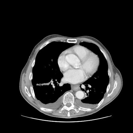
Usually carried out with contrast enhancement.
However, pleural effusions are not entirely innocuous. This should be done before the. There are normally a few milliliters of fluid in the pleural space; Usually carried out with contrast enhancement. It can be estimated, on the basis of if the imaging findings and the analysis of the pleural effusion fluid are inconclusive, pleural biopsy may be needed. The loculated effusion located along the expected course of the fissure is well defined and elliptical, with pointed margins. Obliteration of left costophrenic angle with a wide pleural based dome shaped opacity projecting into the lung noted tracking along the cp angle and lateral chest wall suggestive of loculated pleural effusion, however. Differentiate from an elevated hemidiaphragm. Return back by 'esc' key or x button in the right bottom corner. Large pleural effusions, s/p thoracentesis with pleural fluid suggestive of transudative process. Case contributed by dr prashant mudgal. Pleural effusion, small to moderate 1 of 5 70: Fundamentally a pleural effusion refers to the collection of fluid between the parietal and visceral pleura.
However, patients can also have neutrophilic loculated tpe, although little data are available concerning the incidence and characteristics of this form of tpe. Learn about pleural effusion including causes of pleural effusion. Improved after thoracentesis and diuresis. Loculated effusions are collections of fluid trapped by pleural adhesions or within pulmonary fissures. Ct of the thorax ± abdomen:
Diagnostic Utility And Clinical Application Of Imaging For Pleural Space Infections Chest from els-jbs-prod-cdn.jbs.elsevierhealth.com Pleural effusion 1 of 3 67: Pleural effusions are very common, and physicians of all specialties encounter them. Pleural thickening or attenuation of subcostal fat on ct suggest infection of the pleural cavity intrapleural fibrinolytics in loculated ptb may facilitate pe resolution and reduce residual pleural thickening (>10mm). Most likely secondary to left ventricular diastolic dysfunction. Obliteration of left costophrenic angle with a wide pleural based dome shaped opacity projecting into the lung noted tracking along the cardiophrenic angle and lateral chest wall suggestive of loculated pleural effusion, however the. Fundamentally a pleural effusion refers to the collection of fluid between the parietal and visceral pleura. Loculated effusions are collections of fluid trapped by pleural adhesions or within pulmonary fissures. Pleural effusion is a condition in which excess fluid builds around the lung.
The opacity is effusion is sometimes hard to smoothly marginated and biconvex.
Learn about pleural effusion including causes of pleural effusion. Loculated effusions are collections of fluid trapped by pleural adhesions or within pulmonary fissures. Pleural effusion, small to moderate 2 of 5 71: A pleural effusion is accumulation of excessive fluid in the pleural space, the potential space that surrounds each lung. Ct scans for pleural effusion should be performed with contrast enhancement of the pleura and before complete drainage of pleural fluid. A pleural effusion represents the disruption of the normal mechanisms of formation and drainage of fluid from the pleural space. Pleural thickening or attenuation of subcostal fat on ct suggest infection of the pleural cavity intrapleural fibrinolytics in loculated ptb may facilitate pe resolution and reduce residual pleural thickening (>10mm). Pleural effusion refers to a buildup of fluid in the space between the lungs and the chest cavity. This should be done before the. A rational diagnostic workup, emphasizing the most common causes. It can be estimated, on the basis of if the imaging findings and the analysis of the pleural effusion fluid are inconclusive, pleural biopsy may be needed. Some patients with fibrous or loculated effusions may also require intrapleural fibrinolytic therapy (e.g. In loculated parapneumonic effusions computed tomography (ct).
In loculated parapneumonic effusions computed tomography (ct) loculated pleural effusion. This should be done before the.


0 Komentar